What is Keratoconus?
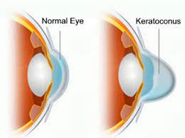
Keratoconus refers to progressive thinning and conical protrusion
of cornea. Cornea is the front most layer of the eye, which is
normally shaped like a mound. This abnormal conical shape of
cornea (ectasia) progressively becomes steeper with localized
corneal thinning, which leads to protrusion of the thinned cornea. This causes blurring of vision
(due to myopia and astigmatism). It usually manifests in the second and third decades of life. It
may be associated with eye allergy and usually affects both the eyes.
What are the symptoms of Keratoconus?
The most common symptom of keratoconus is blurring vision of the affected eye. There can be:
Blurry, double vision
Frequent change of power of glasses (due to change in corneal power)
Distorted images and decreased vision quality due to myopic astigmatism.
Itching and constant desire to rub the eyes (if associated with eye allergy)
In advanced stages, there may be total whitening of cornea with increased
sensitivity to light (acute hydrops stage)
The degree to which vision is affected will depend on the stage of keratoconus, extent of corneal
thinning, the size and degree of conical protrusion in the cornea.
What is the cause of keratoconus ?
While the cause is still unknown, Keratoconus occurs due to a combination of genetic,
environmental and hormonal causes. There is often an underlying eye allergy that aids in
progression of keratoconus. Collagen is an important constituent of corneal tissue. In
keratoconus, disorganization of collagen is seen in cornea.
Is keratoconus common ?
Keratoconus is a relatively common cause of blurred vision among adolescent, with a prevalence
of approximately 1:2000; onset is most common in adolescent age group and certain ethnic
groups such as Middle East African, Asian countries (particularly India, Sri Lanka). Both the eyes
are affected in nearly 80-85% of cases.
Various risk factors for keratoconus are: pubertal age, positive family history of keratoconus,
history of eye rubbing, eye allergy (atopy) or vernal keratconjunctivitis, asthma or hay fever,
connective tissue disorders, floppy eyelid syndrome, retinal hereditary diseases.
How can we diagnose Keratoconus ?
Preliminary diagnosis is by clinical examination with Slit lamp biomicroscopy. Investigations are
done to further confirm keratoconus or to diagnose very early or subtle changes of keratoconus
that are not visible clinically to an eye specialist (i.e. keratoconus suspects). These investigations
are:
• Corneal Topography
• Corneal Pachymetry
• Retinoscopy
How can keratoconus be treated?
There is no 100% cure for keratoconus, only a problem-targetted management approach is
followed. The various options are:
• Spectacles
• Contact lenses- soft and rigid contact lenses, Rose K2 lenses
• Corneal Collagen Crosslinking
• Corneal Transplantation
• Intracorneal ring segments (INTACS)
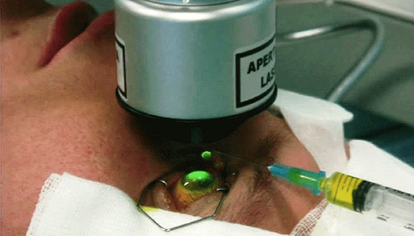
What is the corneal collagen crosslinking (CXL) ?
Corneal collagen crosslinking (CXL) is a surgical procedure, wherein
cornea is treated with riboflavin (Vitamin B2) and ultraviolet (UVA)
radiation directed on corneal surface using a portable machine. CXL treats the corneal disruption by increasing the strength
and rigidity of the corneal tissue by 330% (as per studies).
Step 1 - Corneal epithelium removal
Step 2 - Riboflavin treatment
Step 3 - UVA treatment
Am I suitable candidate for CXL ?
To decide if you are suitable for CXL, eye surgeons usually look for progression of keratoconus by
repeating investigations (corneal topography and pachymetry) at 3 monthly intervals. If criteria of
progression is satisfied, CXL may be done to arrest progression. In some cases, clinicians may go
for CXL if they see a risk of clinical progression.
What are the complications of CXL surgery ?
CXL has a very low of complications. Some patients may experience hazy
vision (7-10%) or develop corneal infection (2-3%) after CXL.
What is the anaesthesia used for this surgery ?
CXL is performed under a topical anaesthetic, which means you will be awake throughout your
operation and only drops will be used to make your eye numb. These drops will prevent you from
feeling any pain during the operation. You will not be able to see details of what is happening, but
you might be aware of the bright lights or movement in the operating theatre. During the operation, we will ask you to lie as flat as possible
and keep your head still.
At the end of the operation, we usually put a pad and shield over your eye to protect it. These will
be removed the morning after your surgery. Some medications will be given in postoperative
period for about 6 weeks.
What are the other surgical options for keratocous?
For keratoconus, if patient is contact lens intolerant or there is advanced disease, surgeons may
advice corneal transplantation procedure (partial or whole). In these, a part or whole of donor
cornea is cut and sutured with the recipient’s cornea, in order to remove the diseased thinned out
cornea with conical protrusion to restore the normal anatomy.
In stable intermediate stages of keratoconus, one has an option of INTACS, i.e placement of
PMMA ring segments into the corneal tissue to increase the strength of the
cornea.
Synergy Eye Care is well equipped and its doctors are well experienced in treating this disease using required procedures and /or surgeries with good results.
Disclaimer: Information published here is for educational purposes only and is not intended to replace medical advice. If you suspect that you have a health problem, please consult your doctor immediately