What is Vitreomacular Traction (VMT)?
The macula is a small area in the centre of the retina where light is sharply focused to produce the
detailed colour vision needed for tasks such as reading and driving.
Vitreous is a clear, gel-like substance that fills the middle of the eye and is firmly attached to the
retina by millions of microscopic fibers. As the eye ages, or as a result of eye disease, the vitreous
shrinks and pulls away from the retina. The vitreous, over time, separates completely from the
retina. This is called a posterior vitreous detachment (PVD) and is usually a normal part of aging.
In some people with PVD, the vitreous doesn’t detach completely. Part of the vitreous remains
stuck to the macula, at the center of the retina. The vitreous pulls and tugs on the macula, causing
vitreomacular traction (VMT)
What causes vitreomacular traction?
VMT is usually caused by part of the vitreous remaining stuck to the macula during a posterior
vitreous detachment.
In healthy eyes, VMT is not common. People with certain eye diseases may be at a higher risk for
VMT, including those with:
• High myopia
• Age-related macular degeneration (AMD)
• Diabetic eye disease
• Retinal vein occlusion
What are the symptoms of VMT?
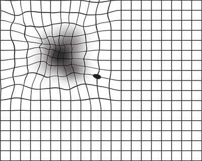
• Distorted vision that makes a grid of straight lines appear wavy, blurry, or blank.
• Seeing flashes of light in your vision
• Seeing objects as smaller than their actual size
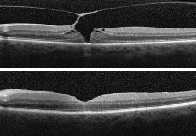
How can we diagnose VMT ?
Optical Coherence Tomography (OCT) helps diagnose VMT. It also helps assess associated
changes in macula like macular edema, epiretinal membrane and macular degeneration.
How can VMT be treated ?
Various treatment options include :
1. Observation (‘wait and watch’ approach)
- If your VMT is mild and not affecting your vision
- Monitor your vision at home each day with an Amsler grid.
2. Surgery
Surgery is the preferred treatment option in cases where VMT is leading to vision threatening retinal conditions, such as:
• Macular hole (when tugging of the vitreous creates a hole in the macula)
• Macular pucker (when macular scar tissue builds up and distorts vision)
• Cystoid macular edema (swelling of the macula)
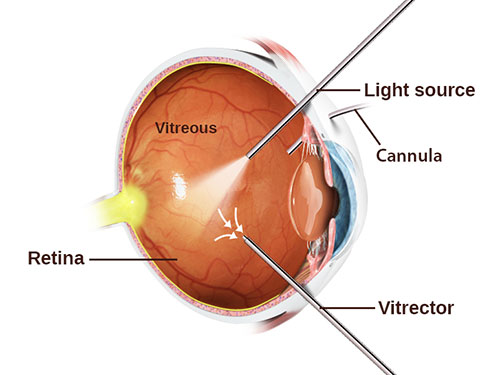
Surgery undertaken for VMT is vitrectomy, to restore
the macula to its normal (lying flat) shape. The surgeon
uses tiny instruments to remove the vitreous from the eye
and replaces it with a saline fluid or air. Any scar tissue on
the macula is also peeled with special instruments under a
microscope. This relieves the traction that is damaging the
macula.
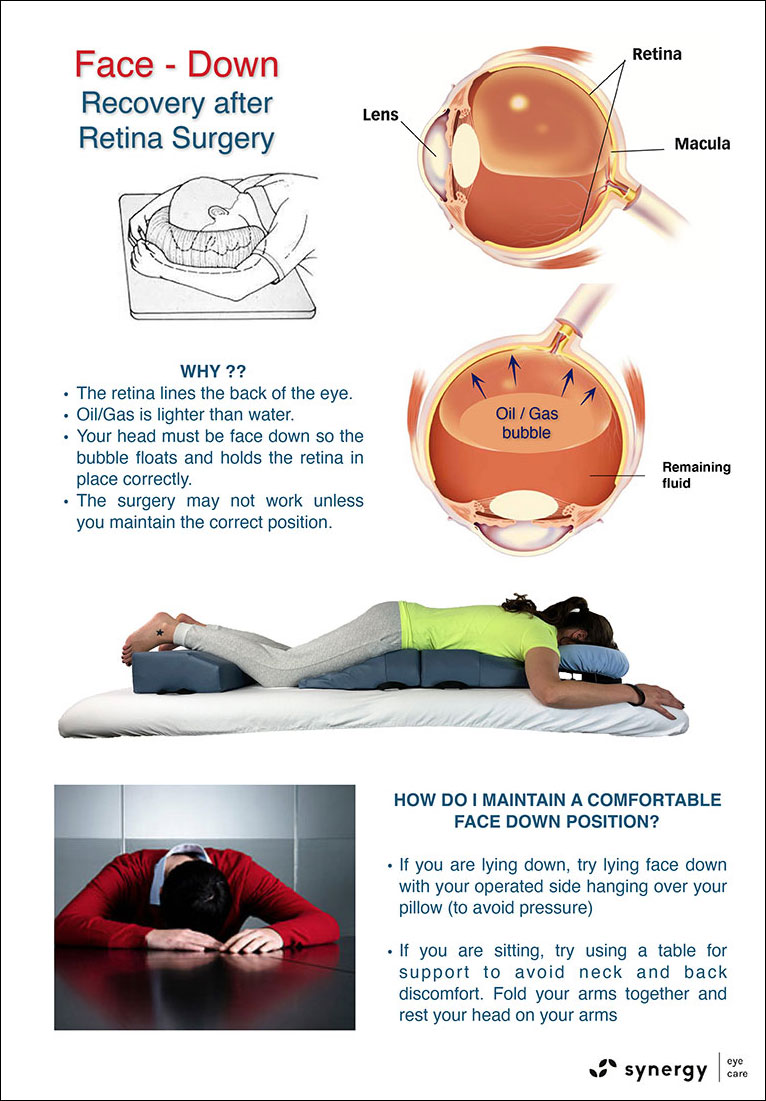
What are the precautions required after vitrectomy surgery ?
Surgery where gas is injected in the eye, the patient may need to maintain a face down position to help push retina back in place. As long as the gas is inside the eye, the vision would be blurred and the patient is not allowed to take air travel.
How much vision recovery to expect after surgery ?
The vision recovery is dependent upon the condition of the retina and the timing of surgery. Generally speaking, earlier surgery leads to better visual results. The vision mostly improves, but may not become as good as totally normal.
Synergy Eye Care is well equipped and its doctors are well experienced in treating this disease using required procedures and /or surgeries with good results.
Disclaimer: Information published here is for educational purposes only and is not intended to replace medical advice. If you suspect that you have a health problem, please consult your doctor immediately