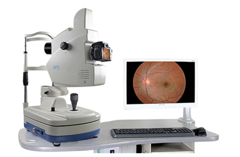
A digital fundus camera is used to take an image of the
fundus — the back portion of the eye that includes the retina,
macula, fovea, optic disc and posterior pole.
The resulting image can then be used by an ophthalmologist
for diagnosis and treatment.
WHAT IS THE PROCEDURE?
Your ophthalmologist will put drops
in your eyes to dilate (widen) your
pupil.
You have to sit in front of the fundus
camera with your chin on a chin rest
(an attachment) and your forehead
against the bar.
Your doctor will focus and align the
fundus camera on you eye.
As soon as the doctor presses the
shutter release, flash fires that
create a photograph of the interior
surface of your eye.
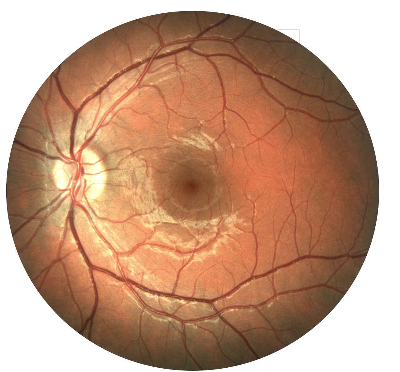
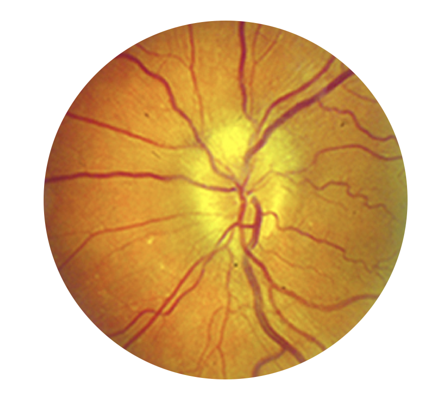
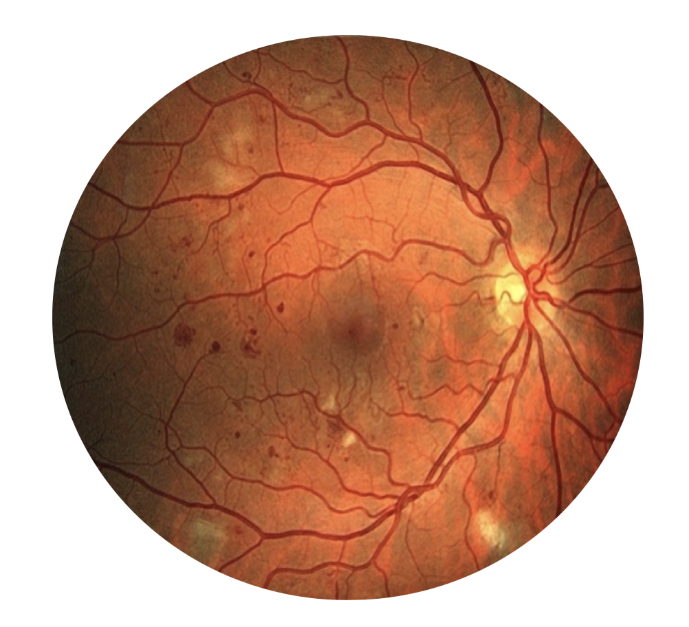
WHY IS FUNDUS PHOTO PERFORMED?
Fundus photo helps detect, treat and
follow various disorders of retina and
optic nerve head including :