Macular Degeneration (ARMD)
The macula is the part of the retina, which provides us
central vision and allows us to see fine detail, such as
recognizing a face, reading, or watching television.
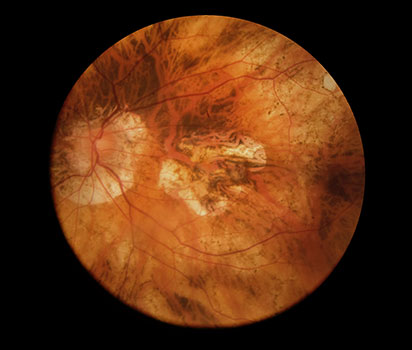
Macular Degeneration is a condition in which the
macula gets damaged. It is often related to aging, and
is commonly referred to as Age-related Macular
Degeneration (AMD). The late stage, associated with
vision loss, is the most common cause of irreversible
blindness in people over the age of 50. It affects the
central vision, especially while reading.
Most often vision loss starts in one eye. Because the
healthy eye compensates for the loss of vision in the
damaged eye, Macular Degeneration may initially go
unnoticed. In many cases it will ultimately affect
vision in the other eye as well.
What are the types of Age-related Macular Degeneration (AMD)?
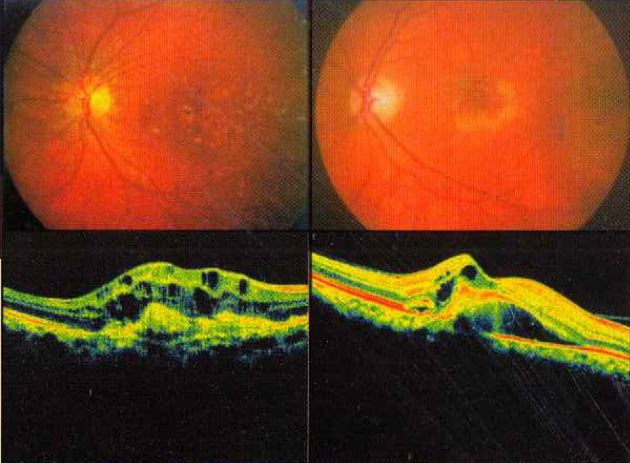
Dry AMD: The retina becomes thinner (atrophic) and
stops functioning. This may cause some people to
detect "blank" areas in their central vision. The vision
loss due to this Dry AMD is not very severe as
compared to the Wet AMD. While there is no
treatment available for people with Dry AMD,
various low vision aids are available to help these
people see well and perform daily activities.
Wet AMD: Abnormal blood vessels grow under the
macula. These abnormal vessels leak fluid and blood,
and thus cause swelling and scar tissue formation,
leading to distorted vision and severe vision loss.
Why is Early Detection important?
The vision lost due to AMD is generally irreversible,
and the treatment methods try to preserve vision but
may not improve vision. Hence it is important to detect
this disease at an early stage, before it has caused
significant vision loss.
How is Macular Degeneration or AMD detected?
In the early stages of AMD, a person's vision may
become blurred or distorted.
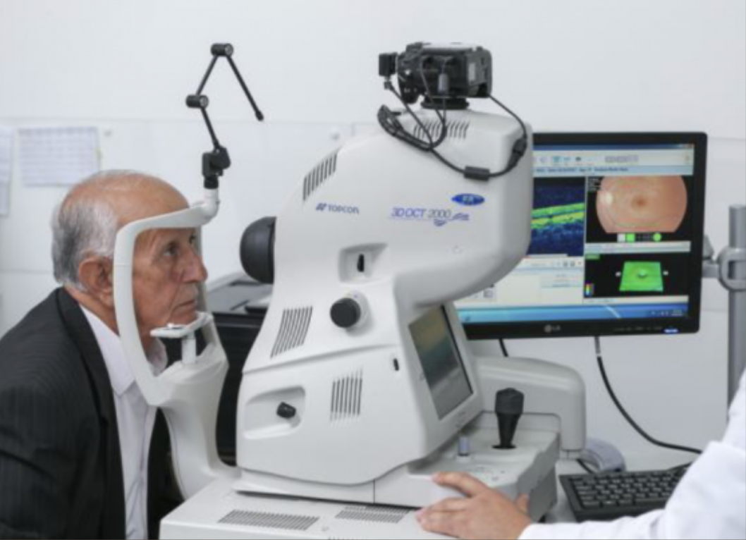
A retinal examination, with
the help of special tests like Fluorescein Angiography,
OCT etc. can help the eye specialist to diagnose the
condition. Since many times the patient may not notice
the initial distortion or blurring of vision, the key to
preventing vision loss due to AMD is regular eye
examinations for patients above 40 years of age. These
regular checkups are also useful in detecting other
potentially serious diseases like Glaucoma.
What are the Treatments available?
Untreated, AMD is known to progress and lead to
further loss of vision, the rate of deterioration being
faster in the wet type. Antioxidants and Multivitamin
capsules may have a role in preventing or decreasing
the speed of progression of the disease. In Wet AMD,
additional methods of treatment are required to arrest or
at least retard the progression of the disease. There have
been many exciting developments in the treatment of
Wet AMD with better results now. The best-suited
treatment modality is decided by the eye specialist after
discussing with the patient. The most popular and
established modes of treatment are:
Intravitreal Injections: This is the exciting new
development in which certain special medicines like
anti-VEGF agents (Lucentis/Accentrix/Razumab,
Eylea, Avastin etc.) are injected in small quantities
within the eye to arrest the disease. The success rate
for maintaining the vision is as high as 90% and in
about 30% of cases, there is even an improvement of
vision. However, the effect of these injections is not
permanent and generally repeated injections (at 4-
6 weeks interval) may be required.
Conventional Laser: Burns the abnormal blood
vessels and thus stops the leakage. However, since it
also damages the normal retina structures, it may
itself lead to decreased vision. Hence, it is suitable
only in selected cases where the new vessels are not
very close to the central macular area.
After advent of Intravitreal injections,
lasers are used in selected cases in
conjunction with intravitreal injections.
More than one sitting of these treatments may be
required. The aim of the treatment is to try to
preserve the vision and not to improve the vision. A
special
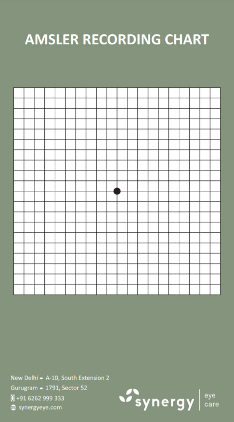
Amsler Grid is given to the patient to
monitor the progress of the disease. With timely and
proper treatment and regular follow ups, majority of
the patients are able to maintain useful vision to
perform their daily activities and lead a socially
productive life.
 Download Amsler Chart pdf
Instructions
- The chart is to be viewed in normal room illumination / light
- Place the chart at normal reading distance (28-38 cms)
- Wear the reading spectacles and view the chart with one eye covered
- Throughout the entire examination keep looking at the central fixation spot. Do not look anywhere else.
- While looking at the central fixation spot. Check whether:
- Is the centre spot visible?
- While viewing the centre black spot,
can you see all four sides?
- Do you see the entire grid intact? Is
any area within the grid not visible?
- Are the horizontal and vertical lines
straight and parallel?
- Are all the squares of equal size and
shape?
- If you notice any blurring or distortion of the lines, or you are unable to see any part of the grid, consult your ophthalmologist immediately.
Synergy Eye Care is well equipped and its doctors are well experienced in treating this disease using required procedures and /or surgeries with good results.
Disclaimer: Information published here is for educational purposes only and is not intended to replace medical advice. If you suspect that you have a health problem, please consult your doctor immediately


