What is Vein Occlusion ?
Vein occlusion is a condition in which the retinal veins that drain blood from the retina closes off
partially/completely.
What are the different types of vein occlusions?
There are two types of RVO:
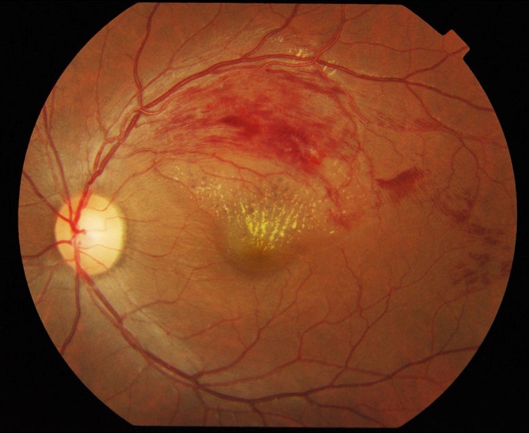
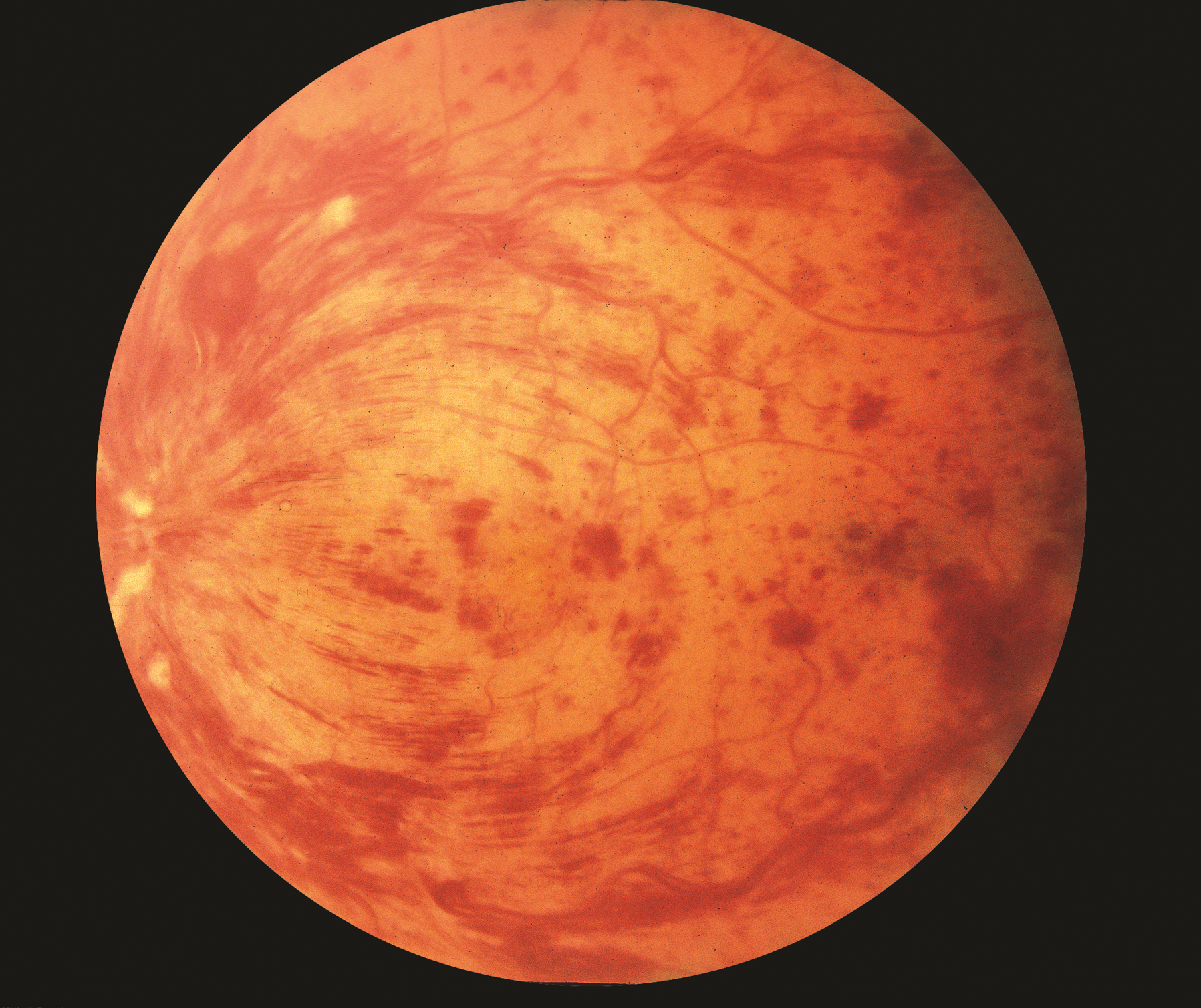
• Central retinal vein occlusion (CRVO) is the blockage of the main retinal vein.
• Branch retinal vein occlusion (BRVO) is the blockage of one of the smaller branch vein
What are the symptoms?
The symptoms of retinal vein occlusion range from subtle to very obvious. There is painless
blurring or loss of vision. It almost always happens in just one eye. At first, the blurring or loss of
vision might be slight, but it gets worse over the next few hours or days. Sometimes there is a
complete loss of vision almost immediately.
What are the Risk factors associated with RVO?
Age - The most important factor. Over 50% of cases occur in patients older than 65.
Hyperlipidaemia is present in one-third or more of patients, irrespective of age.
Diabetes mellitus is present in up to 15% of patients over 50 years of age overall.
Glaucoma and Ocular Hypertension are associated with a higher risk of CRVO and possibly BRVO.
Oral contraceptive pill - In younger females, the contraceptive pill is the most common underlying association.
Smoking may be associated with an increased incidence of RVO.
How to we diagnose vein occlusions?
Ophthalmoscopy: The changes caused by RVO may be seen by examination of the retina
with an instrument called an ophthalmoscope.
Optical Coherence Tomography (OCT): This is a high definition image of the retina taken by
a scanning ophthalmoscope with a resolution of 5 microns. These images can determine the
presence of swelling and edema by measuring the thickness of your retina. The doctor will
use OCT images to objectively document the progress of the disease throughout the course
of your treatment.
Fluorescein angiography: This is a test procedure in which a dye that is injected into a vein
in the arm travels to the retinal blood vessels. Special photographs allow the doctor to see
the vessels.
How is retinal vein occlusion (RVO) treated?
Vision may come back in some eyes that have had a retinal vein occlusion. About 1/3 have some
improvement, about 1/3 stay the same and about 1/3 gradually improve, but it can take a year or
more to learn the final outcome. In some cases, the blocked vessels will lead to fluid
accumulation in the retina, like sponges absorbing water. In others, they may cause the formation
of new blood vessels.
Some of the treatments for retinal vein occlusion include:
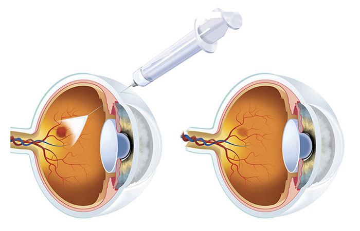
Intravitreal injection of anti-vascular endothelial growth factor (VEGF) drugs: These
drugs target VEGF, which is an important growth factor that causes macular edema.
Intravitreal injection of corticosteroid drugs : These drugs combat the inflammatory
components which lead to edema.
Focal laser therapy : This treatment provides lasers to areas of swelling to cause a
reduction in edema.
Pan-retinal photocoagulation therapy : This treatment is used when patients have new
blood vessel formation following the retinal vein occlusion.
Vitrectomy surgery: The surgery is required sometimes to manage the complications of vascular block like vitreous hemorrhage, retinal detachment etc.
Synergy Eye Care is well equipped and its doctors are well experienced in treating this disease using required procedures and /or surgeries with good results.
Disclaimer: Information published here is for educational purposes only and is not intended to replace medical advice. If you suspect that you have a health problem, please consult your doctor immediately