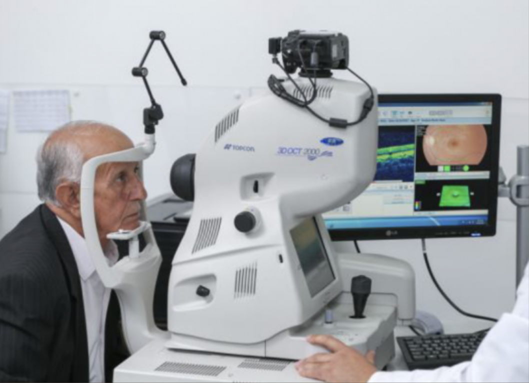

OCT is a non-invasive imaging method used to take scans of anterior surface of eye, the back layer of the eye called the retina and optic nerve. With OCT your ophthalmologist can see each of the retina’s distinctive layers.
Patient will sit in front of the OCT machine and rest head on a support to keep it motionless. The equipment will then scan the eye without touching it. Scanning takes about 2-3 minutes.
WHY IS OCT PERFORMED?OCT Macula is used to evaluate many eye disorders, including:
Macular Edema
Macular Hole
Age-related Macular Degeneration (ARMD)
Vitreo-Macular Traction (VMT)
Central Serous Chorioretinopathy (CSCR)
Epiretinal Membrane
OCT RNFL (Retinal Nerve Fibre
Layer) can help detect changes
caused by glaucoma.
AS-OCT (Anterior Segment OCT)
helps diagnose and treat
disorders of cornea, angle and
anterior chamber.


